〠Instrument R & D 】 The traditional optical microscopy system is limited by Abbe's diffraction limit principle, and it can't distinguish things with a size less than ~ 200nm. In order to break through the diffraction limit, super-resolution fluorescence microscopy technology came into being in the field of biological imaging It is widely used. According to the imaging acquisition process, super-resolution methods can be divided into two categories. One is single-molecule localization microscopy (SMLM), which isolates each luminescent molecule for individual localization through the optical switching characteristics of fluorescent molecules.
This type of method is not limited by the diffraction limit, and can obtain an ultra-high resolution of 10-40 nm, but due to the cycle step of molecular activation bleaching, the acquisition speed and imaging time are slow. The other is super-resolution microscopy technology such as structured light illumination and wide-field imaging, which can achieve a certain degree of response difference between adjacent regions / fluorescent molecules to improve resolution. The method of wide-field imaging has a high time acquisition efficiency, but because all the molecules in the field of view are excited at the same time, its resolution is often above 100 nm. At present, there is still no method that can theoretically effectively take into account the time acquisition efficiency of wide-field imaging and the spatial resolution of single-molecule positioning methods. Therefore, a technical solution for efficiently modulating fluorescent molecules based on wide-field imaging is urgently needed.
The essence of the super-resolution method is to obtain the spatial location of a single fluorescent molecule by identifying its independent emission characteristics. If the fluorescence emission difference of each luminescent molecule within the diffraction limit in wide-field imaging can be actively controlled, it is possible to obtain better super-resolution microscopic results.
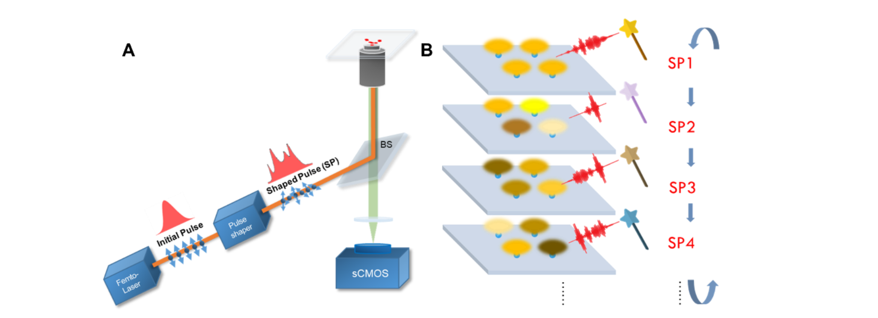
Recently, the extreme optics research team of the State Key Laboratory of Mesoscopic Physics of the School of Physics proposed a method of obtaining super-resolution fluorescence microscopy (SNAC) based on the principle of quantum coherent control to actively modulate molecular fluorescence emission, which achieves resolution under wide-field imaging. Promote. Under the ZnCdS quantum dot system, the research group obtained differential excitation of each quantum dot within the diffraction limit range. By designing multiple shaping pulses, the fluorescence difference of a single ZnCdS quantum dot will be enhanced. The research team periodically changed the shaping pulse and Fourier enhancement to extract the difference in fluorescence response.
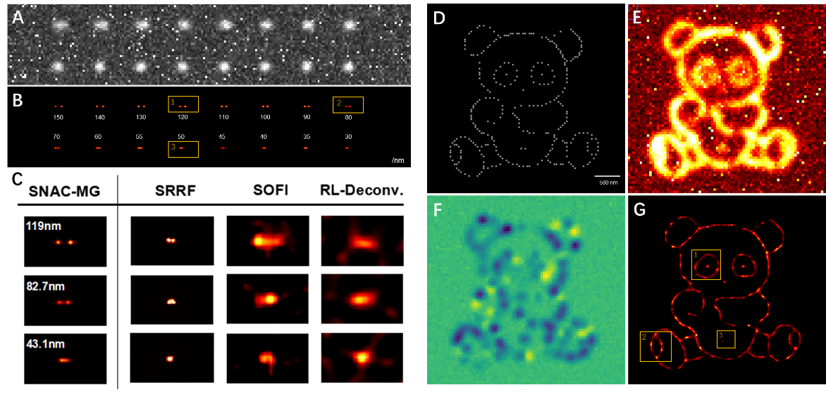
At the same time, the actively controlled image acquisition scheme can effectively suppress the Poisson random noise that does not change with the modulation period in the system and the fixed noise caused by the CMOS process, which greatly improves the signal-to-noise ratio. Then, the research team used the independently developed mixing cycle (Combination-FFT) and multi-Gaussian fitting positioning algorithm to obtain the final super-resolution reconstruction results. The study simulates the super-resolution localization of the adjacent two-point fluorescence emission, and the results can well distinguish the adjacent fluorescent molecules as low as 50nm.
For densely marked linear structures, the resolution of SNAC has also been significantly improved, with a radial positioning accuracy of around 30 nm. In the vascular structure area of ​​the COS7 cell sample marked by quantum dots, the parallel orientation and posture arrangement of the vascular tube and the adjacent double peak of 95.3 nm in the fiber intersection area are clearly observed, showing that there are a variety of wide-field super-resolution methods. Better reconstruction results. This study introduces pulse shaping as a new control dimension to fluorescence super-resolution, and raises the resolution of wide-field super-resolution imaging technology to the level of 50 nm, which is close to the single-molecule localization method.

Figure 1 Experimental device for active control of super-resolution method

Figure 2 Super-resolution simulation verification of active control of adjacent double points and complex two-dimensional structures

Figure 3 QD625 quantum dot labeled COS7 cell biological sample active control super-resolution reconstruction image
The work was published online by Science Advances on April 17, 2020, entitled "Super-resolution nanoscopy by coherent control on nanoparticle emission" (DOI: 10.1126 / sciadv.aaw6579). The first author of the paper is Liu Congyue, a doctoral student of Peking University. Associate Professor Wang Shufeng and Academician Gong Qihuang are the corresponding authors of the paper. This work was supported by the National Natural Science Foundation of China, the Ministry of Science and Technology and the Ministry of Education Nanoelectronics Frontier Innovation Center.
Silicone Baby Bowl,Suction Cup Bowl,Weaning Suction Bowls,Silicone Weaning Bowl
Hubei Daxin Electronic Technology Co., LTD , https://www.nbaiwellgroup.com